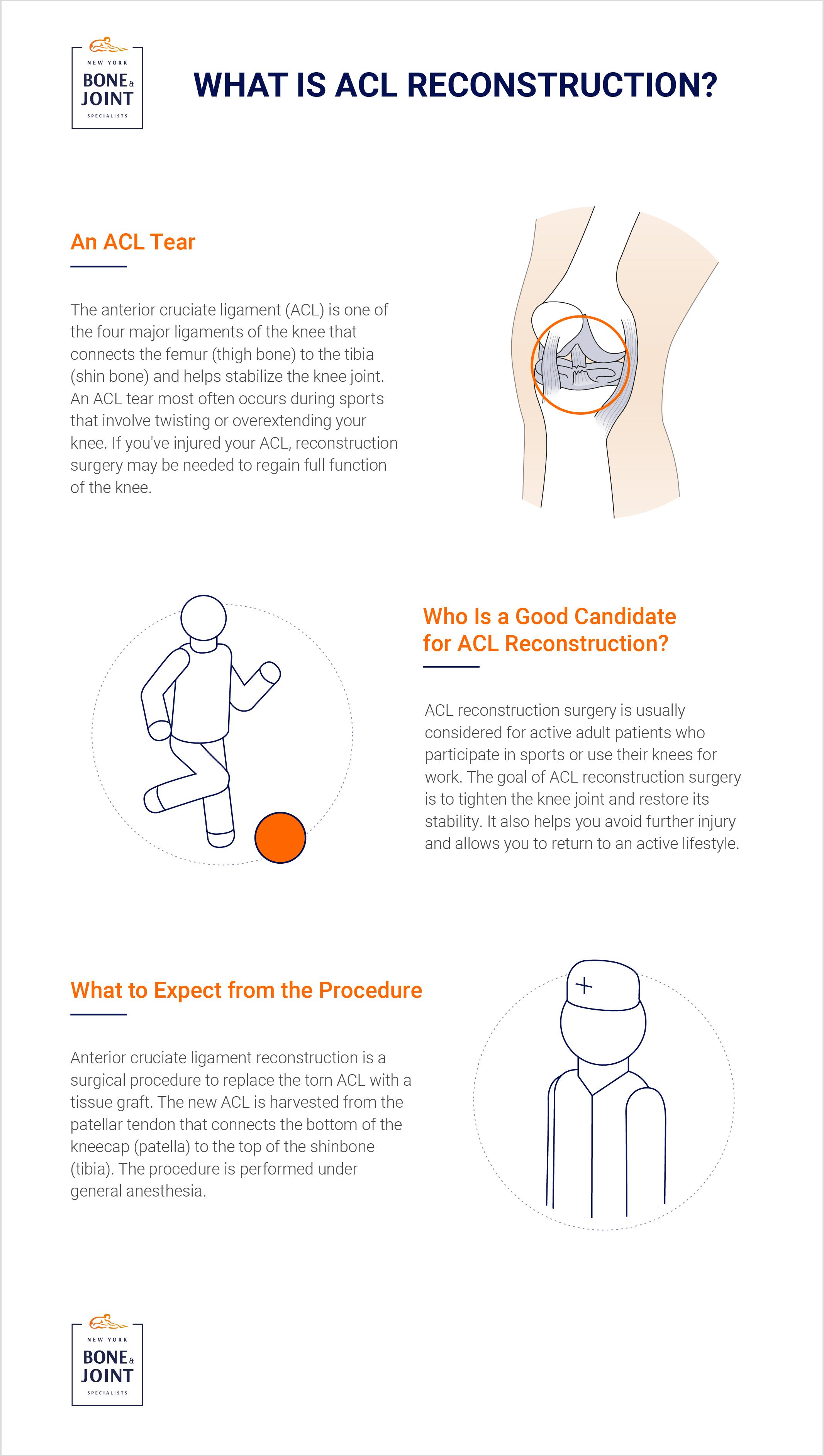
Anterior cruciate ligament (ACL) reconstruction is surgery to reconstruct the torn ligament of your knee with a tissue graft. The anterior cruciate ligament is one of the four major ligaments of the knee that connects the femur (thigh bone) to the tibia (shin bone) and helps stabilize the knee joint. Ligaments are tough, non-stretchable fibers that hold your bones together. Anterior cruciate ligament prevents excessive forward movement of the lower leg bone (tibia) in relation to the thigh bone (femur) as well as limits rotational movements of the knee. An ACL tear is a common knee ligament injury. If you have injured your ACL, surgery may be needed to regain full function of your knee.
CAUSES OF ACL INJURY
An ACL injury most commonly occurs during sports that involve twisting or overextending your knee. The ACL can be injured in several ways:
- Sudden directional change in motion
- Slowing down while running
- Improper landing from a jump
- Direct blow to the side of your knee, such as during a football tackle
SYMPTOMS
When you injure your ACL, you might hear a loud “pop” sound and experience a buckling of the knee. Within a few hours of an ACL injury, your knee may swell due to the bleeding from blood vessels within the torn ligament. You may also notice instability of the knee, especially when trying to change direction of the knee, during movement.
DIAGNOSIS
An ACL injury can be diagnosed with a thorough physical examination of the knee and diagnostic tests such as X-rays, MRI scans and arthroscopy. X-rays may be needed to rule out any fractures. In addition, your doctor will often perform the Lachman’s test to see if the ACL is intact. During a Lachman’s test, knees with a torn ACL may show increased forward movement of the tibia compared to a healthy knee. Pivot shift test is another test to assess ACL tear. During the pivot shift test, if the ACL is torn the tibia will move forward on complete straightening of the knee and as the knee bends past 30° the tibia shifts back into correct place in relation to the femur.
TREATMENT
Treatment options include both non-surgical and surgical methods. If the overall stability of the knee is intact, your doctor may recommend non-surgical methods. Non-surgical treatment consists of rest, ice, compression, and elevation (RICE protocol); all assist in controlling pain and swelling. Physical therapy may be recommended to improve knee motion and strength. A knee brace may be needed to help immobilize your knee.
If left untreated, a torn ACL can lead to early arthritis of the knee.
An ACL reconstruction surgery is considered for active adult patients, who participate in pivoting sports and require knee stability for work.
The goal of ACL reconstruction surgery is to tighten the knee joint and restore its stability. It also helps you avoid further injury and allows you to return to your sport.
PROCEDURE
Anterior cruciate ligament reconstruction patellar tendon is a surgical procedure to replace the torn ACL with part of the patellar tendon taken from the patient’s leg. The new ACL is harvested from the patellar tendon that connects the bottom of the kneecap (patella) to the top of the shinbone (tibia). The procedure is performed under general anesthesia.
Your surgeon will make two small cuts around your knee. An arthroscope, small video camera is inserted through one incision to see the inside of the knee joint. Along with the arthroscope, a sterile solution is pumped into the knee to expand it and enable the surgeon to have a clear view of the inside of the joint. The torn ACL will be removed and the pathway for the new ACL is prepared.
Your surgeon makes an incision over the patellar tendon and takes out the middle third of the patellar tendon, along with its bone attachment on each end. The remaining portions of the patellar tendon on either side of the graft are sutured back after its removal. Then the incision is closed. The arthroscope is reinserted into the knee joint through one of the small incisions. Small holes are drilled into the upper and lower leg bones where these bones come together at the knee joint. The holes form tunnels in your bone to accept the new graft. Then the graft is pulled through the predrilled holes in the tibia and femur. The new tendon is then fixed into the bone with screws to hold it into place while the ligament heals into the bone. The wounds are then closed with sutures and a dressing is placed.
_________________________________
EXPERIENCING PAIN? DO YOU HAVE AN INJURY?
Our Specialists are here to help.
Book an appointment with NYC’s best orthopedic specialists to discuss your condition. Fill out the form below and you will receive a call from our office within 5-10 minutes. We’ll book an appointment at a time and location that work for you, and send you a reminder by email.